What is the sulcus terminalis
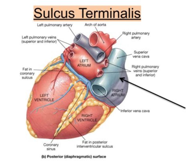
The sulcus terminalis is an inconsistent depression of the myocardial surface of the right atrium of the heart. It extends a short distance inferiorly and to the right from the space intermediate to the superior vena cava and right auricle.
What is the function of sulcus terminalis in tongue?
Upper surface of the tongue The terminal sulcus is a shallow groove that runs forward as a shallow groove in a V shape from the foramen cecum, forwards and outwards to the margins (borders) of the tongue. The terminal sulcus divides the tongue into a posterior pharyngeal part and an anterior oral part.
What is terminal crest in the right atrium?
Right atrium The terminal crest (or crista terminalis) marks the border between the smooth intercaval posterior wall of the RA and the appendage. 3,4. It delineates the border between the smooth wall of the venous component and the rough wall of the appendage (Fig.
Is crista terminalis same as sulcus terminalis?
Crista terminalisFMA9236Anatomical terminologyWhat is the definition of a sulcus?
Definition of sulcus : furrow, groove especially : a shallow furrow on the surface of the brain separating adjacent convolutions.
What is an appendage of the heart?
The left atrial appendage (LAA) is a small, ear-shaped sac in the muscle wall of the left atrium (top left chamber of the heart).
What is the dorsum of the tongue?
The upper surface of the tongue is called the dorsum, and is divided by a groove into symmetrical halves by the median sulcus. The foramen cecum marks the end of this division (at about 2.5 cm from the root of the tongue) and the beginning of the terminal sulcus.
What is Koch's triangle?
Koch’s triangle, named after the German pathologist and cardiologist Walter Karl Koch, is an anatomical area located in the superficial paraseptal endocardium of the right atrium, which its boundaries are the coronary sinus orifice, tendon of Todaro, and septal leaflet of the right atrioventricular valve.What is Coumadin Ridge?
The coumadin ridge is a prominent, muscular ridge of tissue that lies in the left atrium in between the left superior pulmonary vein and the left atrial appendage[2]. It may often appear to be attached to the roof of the left atrial appendage, with a rounded end extending into the left atrium[2].
Does left atrium have pectinate muscles?The pectinate muscles are “teeth of a comb” shaped parallel muscular columns that are present on the inner wall of the right and left atria. The right atrium has thick and coarse pectinate muscles while these are few smooth and thinner in the left atrium.
Article first time published onWhat 3 vessels fill the right atrium?
The blood vessels include the superior and inferior vena cava. These bring blood from the body to the right atrium. Next is the pulmonary artery that carries blood from the right ventricle to the lungs.
What is a Chiari network in right atrium?
Introduction. The Chiari network, encountered infrequently in the right atrium, is a fenestrated, net-like embryonic remnants of valves of sinus venosus, lying closely in relation to the inferior vena cava and coronary sinus, sometimes connecting these with other right atrial structures [1].
What do you mean by sulci and gyri of brain?
Gyri and sulci are the folds and indentations in the brain that give it its wrinkled appearance. Gyri (singular: gyrus) are the folds or bumps in the brain and sulci (singular: sulcus) are the indentations or grooves. … The medial longitudinal fissure is the sulcus that separates the left and right brain hemispheres.
Where is the sulcus located?
Central sulcusLocationCerebral cortexIdentifiersLatinsulcus centralis cerebriNeuroNames48
What does the medulla oblongata mean?
medulla oblongata, also called medulla, the lowest part of the brain and the lowest portion of the brainstem. … The medulla oblongata plays a critical role in transmitting signals between the spinal cord and the higher parts of the brain and in controlling autonomic activities, such as heartbeat and respiration.
What is the function of dorsum?
The top surface, or dorsum, contains numerous projections of the mucous membrane called papillae. They contain taste buds, which are sensitive to chemical constituents of food, and serous glands that secrete some of the fluid in saliva, a substance that moistens the oral cavity and helps lubricate food particles.…
What is the dorsum of the foot?
The dorsum of foot is the area facing upwards while standing. This definition incorporates text from the wikipedia website – Wikipedia: The free encyclopedia. ( 2004, July 22).
What covers the dorsum of the tongue?
The dorsal surface of the tongue is covered with four types of papillae. Filiform are the most numerous papillae and cover the anterior two-thirds of the dorsum of the tongue.
What is the purpose of LAA?
This procedure, called left atrial appendage closure (LAAC), helps prevent stroke by sealing off a small, unnecessary section of the heart called the left atrial appendage (LAA). For people with Afib, most strokes get their start in the LAA because that is where blood clots tend to form.
What does the LAA do?
As the heart pumps, blood travels through the atria and the ventricles. The LAA is hollow, so it fills with blood when the left atrium receives blood and it empties when blood travels out of the left atrium. In most people the left atrial appendage is of little or no concern.
What is the function of the appendage?
1 Introduction. Animal appendages are external projections from the body wall that are used for very diverse functions including locomotion, grooming, and feeding.
What is Coumadin ridge on Echo?
Coumadin ridge is a normal tissue/pericardial folding separating openings of LA appendage and the left upper pulmonary vein on the left lateral atrial wall. Its presence indicates normal opening of left upper pulmonary vein into left atrium thus ruling out TAPVC.
What is a ridge on the heart?
Background: The left atrial ridge is a structure located in the left atrium between the left-sided pulmonary veins ostia and the orifice of the left atrial appendage. Since it was commonly misdiagnosed as a thrombus, the ridge is also known as the “coumadin” or “warfarin” ridge.
What is Marshall ligament?
The ligament of Marshall (LOM) is located on the epicardium between the left atrial appendage and the left pulmonary veins. The corresponding endocardial structure is the left lateral ridge. … These muscle structures can serve as a source of triggers and drivers for AF, and may form the substrates of reentry.
What is the sinus Venarum?
The posterior part of the right atrium is termed the sinus venarum; also, it includes most of the lateral wall of the chamber. It has a relatively smooth surface compared to the anterior part. The posterior and anterior walls merge at the crista terminalis.
Why is triangle of Koch important?
Koch’s triangle is an important area of human heart, which is located in the superficial paraseptal endocardium of the right atrium, used as an anatomical landmark to locate the atrioventricular (AV) node.
Does coronary sinus have valves?
Valve of the coronary sinusTA24030FMA9242Anatomical terminology
What forms the apex of heart?
The apex (the most inferior, anterior, and lateral part as the heart lies in situ) is located on the midclavicular line, in the fifth intercostal space. It is formed by the left ventricle. The base of the heart, the posterior part, is formed by both atria, but mainly the left.
Which heart sulcus contains the great cardiac vein?
The great cardiac vein is the longest venous vessel of the heart; in the majority of our cases it originated at the lower third of the anterior interventricular sulcus (58%).
What is limbus left atrial appendage?
The left atrial appendage (LAA) is a “dog ear”–shaped extension of the LA located along the lateral aspect of the chamber near the MV annulus.
What is the largest artery in the body?
Aorta Anatomy The aorta is the large artery that carries oxygen-rich blood from the left ventricle of the heart to other parts of the body.
