What is the role of Janus Green B stain in the slide preparation
Janus Green B is a basic dye and vital stain used in histology. It is also used to stain mitochondria supravitally, as was introduced by Leonor Michaelis in 1900. The indicator Janus Green B changes colour according to the amount of oxygen present. When oxygen is present, the indicator oxidizes to a blue colour.
What structures does Janus Green B stain in a cell how does it work?
Janus Green B is chemically diethyl safranin-azo-dimethyl aniline. It is oxidized to become colored. The cytochrome c oxidase helps to maintain the dye in an oxidised state in the mitochondria. In its oxidized state it is bluish green in colour.
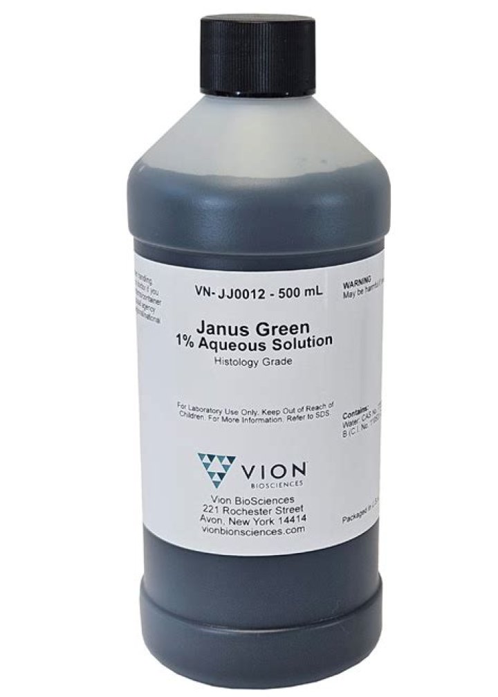
How do you prepare Janus green B solution?
To prepare a Janus Green B solution, use a 1% aqueous solution. Add 1 g of Janus Green B to 75 ml of DI water. Dilute to a final volume of 100 ml with DI water.
What is the role of Janus green B Indicator in anaerobic experiment with yeast?
Answer: The indicator Janus Green B changes colour according to the amount of oxygen present. … In the absence of oxygen, the indicator is reduced and changes to a pink colour.What is the meaning of Janus Green?
Definition of Janus green : a basic azine dye used especially as a vital biological stain (as for mitochondria)
Why does Janus green identify mitochondria?
(d) Janus green is used to stain mitochondria. Janus green act as an indicator and changes colour according to the amount of oxygen present. It oxidizes to blue colour in. presence of oxygen and in its absence changes its colour to pink.
What does Janus green stain?
It has been used to stain biomolecules, nucleic acids and chromosomes. In addition, it is also used to stain various living organisms including fungi, yeast cell, brain, spinal cord, sperms and tissue culture monolayer. Janus Green B acts as an antimalarial agent.
What is Green B?
B sustainable, today. On the occasion of Earth Day, Benetton Group presents GREEN B, the project that rounds up all the brand’s initiatives for sustainability. “GREEN B is the ambassador of Benetton’s innate green soul, the symbol of everything that is sustainable in us.” …Why do we use Diazine green or Janus Green B to glucose solution in the experiment of anaerobic respiration with yeast?
Diazene Green solution is added to the Glucose solution in anaerobic respiration experiment to check the presence of oxygen in glucose solution.
What is used to stain mitochondria?DASPEI (2-(4-(Dimethylamino)styryl)-N-ethylpyridinium iodide) is used to stain mitochondria in live cells. DASPEI (2-(4-(Dimethylamino)styryl)-N-ethylpyridinium iodide) is used to stain mitochondria in live cells.
Article first time published onWhich is vital stain?
Janus green, methylene blue and neutral red are called vital stains.
What is the use of safranin solution?
Safranin is used as a counterstain in some staining protocols, colouring cell nuclei red. This is the classic counterstain in both Gram stains and endospore staining. It can also be used for the detection of cartilage, mucin and mast cell granules.
What is vital staining techniques?
vital staining A technique in which a harmless dye is used to stain living tissue for microscopical observation. The stain may be injected into a living animal and the stained tissue removed and examined (intravital staining) or the living tissue may be removed directly and subsequently stained (supravital staining).
Who is Janus in Greek mythology?
Some scholars regard Janus as the god of all beginnings and believe that his association with doorways is derivative. He was invoked as the first of any gods in regular liturgies. The beginning of the day, month, and year, both calendrical and agricultural, were sacred to him.
What does fast green stain?
fast green A green dye used in optical microscopy that stains cellulose, cytoplasm, collagen, and mucus green. It is frequently used to stain plant tissues, with safranin as a counterstain.
What is ultrastructure of mitochondria?
Ultrastructure of Mitochondria: Mitochondria are bounded by an envelope consisting of two concentric membranes, the outer and inner membranes. The space between the two membranes is called inter-membrane space. A number of invaginations occur in the inner membrane; they are called cristae (Fig. 2.21).
Why basic dyes are often used in microbiology?
Basic Dyes carry a positive charge & are more used for staining than Acidic dyes. This is because opposite charges attract, basic dyes stain the negatively charged components of cells including nucleic acid & many proteins.
Who discovered mitochondria?
Mitochondria, often referred to as the “powerhouses of the cell”, were first discovered in 1857 by physiologist Albert von Kolliker, and later coined “bioblasts” (life germs) by Richard Altman in 1886. The organelles were then renamed “mitochondria” by Carl Benda twelve years later.
Who first introduced the term mitochondria?
The term “mitochondria” was coined in 1898 by Carl Benda, one of the first microbiologists to use a microscope in studying the interior structure of cells.
How does mitochondria power the cell?
Known as the “powerhouses of the cell,” mitochondria produce the energy necessary for the cell’s survival and functioning. Through a series of chemical reactions, mitochondria break down glucose into an energy molecule known as adenosine triphosphate (ATP), which is used to fuel various other cellular processes.
What is the importance of Diazine green?
Diazine green is added to glucose solution to know whether oxygen is present or not in glucose solution. When the availability of oxygen is less than the diazine green changes to pink colour.
What is the use of adding Diazine green to glucose solution what change you notice in glucose solution?
Diazine green solution is used as a test to determine the presence of oxygen in a solution. When the solution is deficient in oxygen then diazine green solution turns it into a pink coloured solution. This confirms the absence of oxygen in the solution. The same thing happens with the glucose solution.
Why lime water is used in this experiment?
Freshly prepared lime water is used in the experiment to test the presence of CO2 because lime water turns milky when CO2 passes through it.
What color do mitochondria stain?
On histological slides stained with hematoxylin and eosin (H&E), the color of cytoplasm in cells containing many mitochondria is: red or pink.
What staining process is used to stain the nucleus mitochondria and chloroplast?
What is Cellular Staining? Cell staining is a technique that can be used to better visualize cells and cell components under a microscope. By using different stains, one can preferentially stain certain cell components, such as a nucleus or a cell wall, or the entire cell.
Which of the following is used to stain the cell?
Methylene blue is used to stain animal cells, such as human cheek cells, to make their nuclei more observable.
What are the purpose of staining?
The main purpose of staining is to highlight cells and parts of cells. Over 20 different types of stains exist, and the type of stain you use depends on what you are looking for.
What is vital and Supravital staining?
A vital stain in a casual usage may mean a stain that can be applied on living cells without killing them. … In supravital staining, living cells have been removed from an organism, whereas intravital staining is done by injecting or otherwise introducing the stain into the body.
What is principle stain?
The basic principle of gram staining involves the ability of the bacterial cell wall to retain the crystal violet dye during solvent treatment. Gram-positive microorganisms have higher peptidoglycan content, whereas gram-negative organisms have higher lipid content.
What is malachite green stain used for?
Malachite Green is used for bacterial spore staining by Schaeffer and Fulton’s method. It can also be used as a simple stain for bacterial cells and in place of methyl-green in Pappenheim stain, when combined with Gram stain.
What is safranin function?
Safranin (also Safranin O or basic red 2) is a biological stain used in histology and cytology. Safranin is used as a counterstain in some staining protocols, colouring cell nuclei red. … It can also be used for the detection of cartilage, mucin and mast cell granules.
