What is a lateral cephalometric radiograph
A lateral cephalometric radiograph (LCR) is a standardised, reproducible radiograph used primarily for orthodontic diagnosis and treatment planning. It is taken from a distance of 1.5m with the head at a right angle to the X-ray
What is cephalometric radiographic method and why it is used by orthodontists?
Cephalometric radiographs A cephalometric radiograph is a radiograph of the head which is taken in a Cephalometer. The Cephalostat is a head-holding device which was introduced in 1931 by Holly Broadbent Sr. The Cephalometer is used to obtain standardized craniofacial images which are available on radiographic films.
What is the difference between panoramic and cephalometric?
Cephalometric Analysis is an X-ray similar to a panoramic X-ray, in that it has the capability of capturing a full view of your skull and neck. A difference is that it is captured using a side-to-side sweeping motion, instead of the full 360 degree non-stop motion used in panoramic X-rays.
What is a lateral Ceph?
A Lateral Cephalogram (or Lat Ceph) is an x-ray taken of the side of the face with very precise positioning so that various measurements can be made to determine the current and future relationship of the top and bottom jaw (maxilla and mandible) and therefore assess the nature of a patient’s bite.What is the meaning of cephalometric?
Cephalometry is the study and measurement of the head, usually the human head, especially by medical imaging such as radiography. … Cephalometry also has a history in phrenology, which is the study of personality and character as well as physiognomy, which is the study of facial features.
How do you take lateral cephalometric radiograph?
A lateral cephalometric radiograph (LCR) is a standardised, reproducible radiograph used primarily for orthodontic diagnosis and treatment planning. It is taken from a distance of 1.5m with the head at a right angle to the X-ray beam at a distance of 30cm, (although this has been found to vary slightly).
What does a cephalometric radiograph show?
Cephalometric x-rays (also called ceph x-rays or radiographs) show a side view of your head, exposing teeth, jaw, and surrounding structures. … This specific type of x-ray is used in diagnosis and treatment planning.
What are lateral oblique radiographs used for?
The lateral oblique x-ray view of the mandible and maxilla taken on an extra-oral film is a frequently used method for giving a record of the teeth in the buccal segments from canine to third molar show- ing the teeth both erupted and unerupted or to assess the positions of unerupted third permanent molars.How is cephalometric measured?
Traditionally, cephalometric analysis has been carried out using a manual method. An acetate sheet is placed over the radiograph and measurements are recorded of the distances and angles between cephalometric landmarks with a ruler and a protractor.
What is cephalic radiograph?A Cephalometric x-ray (CEPH x-ray) consists of a lateral/side-view x-ray of the face – also referred to as Lat CEPH. As with an OPG X-ray a CEPH radiograph is used to study facial bone and soft tissue landmarks and is used in patients being considered for orthodontic treatment.
Article first time published onWhat is a panorex used for?
A panorex is an x–ray that provides a full view of the upper and lower jaws, teeth, temporomandibular joints (TMJs) and sinuses.
What is a panoramic detector?
Panoramic imaging is a novel adaptation of dental radiography applied to show all the patient’s teeth in one large image. … A TDI image sensor has many vertical rows of pixels, which can enhance the signal-to-noise ratio (SNR) and overall image quality by repetitively sampling the image row by row.
What is a Cephalogram used for?
The lateral cephalogram is a profile x-ray of the skull and soft tissues and is used to assess the relation of the teeth in the jaws, the relation of the jaws to the skull and the relation of the soft tissues to the teeth and jaws.
How do you do cephalometric tracing?
Manual tracing of cephalometric Alms is performed by identifying radio-graphic landmarks on acetate overlays and using these reference points to construct lines, planes and angles to enable the measurement of linear and angular values, using a millimetre scale and a protractor.
Why cephalometric analysis is the preferred tool in the diagnosis and treatment of malocclusion?
Cephalometric radiography is an essential tool in the diagnosis and treatment of dental malocclusions and underlying skeletal discrepancies. The use of serial cephalometric radiographs makes it possible to study and predict growth, orthodontic treatment progress and surgical outcome of dentofacial deformity treatment.
What is the importance of Cephalometrics?
The cephalometric roentgenograph has provided a means of accurately appraising the relationships of the parts of the face leading to a description of the mean or average facial form of normal occlusion. It also shows the range of variation that may occur. These abilities permit the attempt to classify facial types.
What is ceramic braces for teeth?
Ceramic braces were the first to offer a much more discreet shape and color than conventional ones. So, they revolutionized the world of orthodontics. These are made with materials very similar to porcelain. Ceramic brackets have a lower resistance to metal brackets, so some people consider them a bit fragile.
What radiograph is used for orthodontic treatment?
A cephalometric X-ray, which is also sometimes referred to simply as a ceph, is a diagnostic radiograph used primarily for orthodontic treatment planning1 . A cephalometric X-ray is taken during the orthodontic records appointment.
What is lateral oblique?
Introduction. Oblique lateral radiographs are extraoral views of the jaws that can be taken using a dental X-ray set (see Fig. 12.1). Before the development of panoramic equipment they were the routine extraoral radiographs used both in hospitals and in general practice.
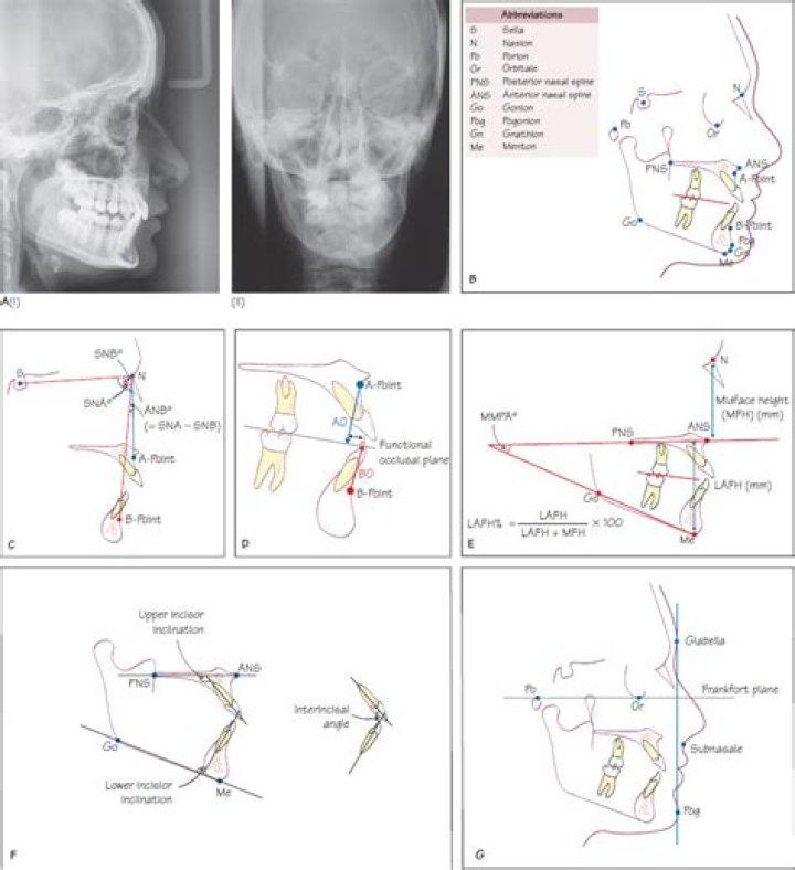
What is the cephalometric measurement used to evaluate the chin position relative to Frankfurt horizontal?
B-point is the most anterior measure point of the mandibular apical base. This angle expresses the horizontal position of the mandible relative to the cranial base using B-point as a cephalometric landmark. This angle represents the extent of chin prominence relative to the reference plane SN.
What is a lateral radiograph?
The lateral chest radiograph is a valuable source of information that has become increasingly undervalued in the era of chest computed tomography. Optimal use of the lateral radiograph requires systematic analysis. First is an overview, followed by analysis of the airway and major hilar structures.
What is an oblique view in radiology?
Oblique – Projection taken with the central ray at an angle to any of the body planes. Described by the angle of obliquity and the portion of the body the X-ray beam exits; right or left and posterior or anterior. For example, a 45 degree Right Anterior Oblique of the Cervical Spine.
Which type of dental radiograph shows 3d images?
Cone beam CT is a type of X-ray that creates 3-D images of dental structures, soft tissue, nerves and bone. It helps guide tooth implant placement and evaluates cysts and tumors in the mouth and face. It also can see problems in the gums, roots of teeth and the jaws.
When should I take panorex?
Panorex x-rays are performed yearly if there are no oral problems. You can have them anytime you want. It depends upon the situation of your teeth. Maybe the teeth ache so much that you feel a dire need of panorex.
What are the three types of dental images?
There are three types of diagnostic radiographs taken in today’s dental offices — periapical (also known as intraoral or wall-mounted), panoramic, and cephalometric. Periapical radiographs are probably the most familiar, with images of a few teeth at a time captured on small film cards inserted in the mouth.
How do you read a panoramic radiograph?
- Describe the location of the lesion.
- Describe the internal structure of the lesion: radiopaque or radiolucent.
- Describe the size, shape and border of the lesion.
- Describe the effect of the lesion to the surrounding structures.
Which pencil is used for cephalometric tracing?
Tracings are used to facilitate cephalometric analysis, as well as in superimpositions, to evaluate treatment and growth changes. Historically, tracings of the cephalometric radiographs are done on an 0.003 inch thick matte acetate paper by using a #3 pencil.
What is Steiner analysis?
The Steiner numerical analysis, which was developed in the 1950s (7–9) suggests a series of measurements not only to diagnose the problem but it also provides guidelines for treatment planning based on the pre- diction of changes that take place as a result of growth and/or orthodontic therapy.
What is orthodontic wire?
An archwire in orthodontics is a wire conforming to the alveolar or dental arch that can be used with dental braces as a source of force in correcting irregularities in the position of the teeth. An archwire can also be used to maintain existing dental positions; in this case it has a retentive purpose.
