What does Magnocellular mean
Magnocellular cells make up the magnocellular layers of the lateral geniculate nucleus. They are relatively large cells that display specialization in detecting aspects of movement, such as the location, speed, and direction of a moving object.
What do Magnocellular cells do?
Magnocellular cells make up the magnocellular layers of the lateral geniculate nucleus. They are relatively large cells that display specialization in detecting aspects of movement, such as the location, speed, and direction of a moving object.
What are M and P cells?
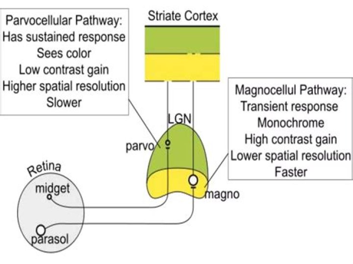
M and P cells also differ in ways that are not so obviously related to their morphology. M cells respond transiently to the presentation of visual stimuli, while P cells respond in a sustained fashion. Moreover, P ganglion cells can transmit information about color, whereas M cells cannot.
What is the difference between the magnocellular and parvocellular pathways?
The Magnocellular pathway carries information about large, fast things (low spatial frequency; high temporal frequency) and is colorblind. The Parvocellular pathway carries information about small, slow, colorful things (high spatial frequency information; low temporal frequency information).Where does the magnocellular pathway go?
The magnocellular pathway is one of the three primary subcortical pathways (magnocellular, parvocellular, and koniocellular pathways) leading from the retina to visual cortex via the lateral geniculate nucleus (LGN).
What is magnocellular theory of dyslexia?
The theory postulates that the magnocellular pathway is selectively disrupted in certain dyslexic individuals, leading to deficiencies in visual processing, and, via the posterior parietal cortex, to abnormal binocular control and visuospatial attention (Stein and Walsh, 1997; Hari et al., 2001).
Is magnocellular faster than parvocellular?
Signals relayed through the magnocellular layers of the LGN travel on axons with faster conduction speeds than those relayed through the parvocellular layers.
What are Parvo and Magno cells?
Magno cells are large, have thick axons and usually collect input from many retinal cells. Parvo cells are smaller, with fine axons and less myelin than mango cells. Konio cells are diverse small cells with wide fields of input consisting of different cells types. The three cellular pathways also differ in function.What are blobs and Interblobs?
Blobs are areas within V1 sensitive to color, whereas interblobs are areas sensitive to the orientation of an object. The interblob cells respond as the simple cells that we have described above. The blobs show color responses, and the layer 4B respond well to moving stimuli and stimuli of very low contrast.
What is striate cortex?The striate cortex is the part of the visual cortex that is involved in processing visual information. The striate cortex is the first cortical visual area that receives input from the lateral geniculate nucleus in the thalamus.
Article first time published onWhat is the magnocellular stream?
The magnocellular visual stream signals us to an awareness of the time properties of objects. For instance, detection of the movement, distance, and speed of an object moving towards us. Small, ‘Parvocellular’ cells. ‘Parvocells’ or P-cells carry visual information along the ventral stream of the brain.
What do M type ganglion cells do?
In addition to being larger themselves, type M ganglion cells have larger receptive fields, propagate action potentials more quickly in the optic nerve, and are more sensitive to low-contrast stimuli.
Where are midget cells found?
Anatomical terminology A midget cell is one type of retinal ganglion cell (RGC). Midget cells originate in the ganglion cell layer of the retina, and project to the parvocellular layers of the lateral geniculate nucleus (LGN).
What visual information would be lost if the optic chiasm were cut?
Damage at site #3: the optic chiasm would be damaged. In this case, the temporal (lateral) portions of the visual field would be lost. The crossing fibers are cut in this example.
Which type of deficit suggests dyslexics have a problem with magnocellular function?
Visual magnocellular deficit theory suggests that the difficulties in the visual processing of dyslexia are caused by the dysfunction of the magnocellular system. However, some researchers have pointed out that previous studies supporting the magnocellular theory did not control for the role of “noise”.
What causes developmental dyslexia?
Dyslexia risk factors include: A family history of dyslexia or other learning disabilities. Premature birth or low birth weight. Exposure during pregnancy to nicotine, drugs, alcohol or infection that may alter brain development in the fetus.
What is rapid auditory processing?
Tallal’s theory asserts that performance on the ART (the measure of rapid auditory processing) predicts reading ability through its influence on phonological representations, which in turn determine phonological awareness and phonological processing ability.
What is color blob?
From Wikipedia, the free encyclopedia. Blobs are sections of the visual cortex where groups of neurons that are sensitive to color assemble in cylindrical shapes. They were first identified in 1979 by Margaret Wong-Riley when she used a cytochrome oxidase stain, from which they get their name.
What are cells in V4 sensitive to?
Visual Area Four (V4, extrastriate cortex) V4 receives information from V2 and is part of the ventral processing stream. Cells in V4 are very responsive to color.
What does the superior colliculus do?
The superior colliculus (SC) is a multisensory midbrain structure that integrates visual, auditory, and somatosensory spatial information to initiate orienting movements of the eyes and head toward salient objects in space.
What are Parvocellular cells?
AKA P-cells. Parvocellular cells make up the parvocellular layers of the lateral geniculate nucleus. They are relatively small compared to magnocellular cells and are important for spatial resolution, visual acuity, and the detailed analysis of shape, size, and color.
What is V4 in the brain?
V4 is the third cortical area in the ventral stream, receiving strong feedforward input from V2 and sending strong connections to the PIT. It also receives direct input from V1, especially for central space. In addition, it has weaker connections to V5 and dorsal prelunate gyrus (DP).
What does the prefrontal lobe do?
The prefrontal cortex performs functions of cognitive control, and is prominently – though not exclusively – involved in working memory organization via central executive processes.
What is visual stria?
The term stria of Gennari is a myeloarchitectural term denoting a greatly thickened outer band of Baillarger (a dense horizontal plexus of myelinated fibers in the internal granular layer (IV) of cerebral cortex ). It is visible to the naked eye and gives the primary visual cortex its name.
Is Akinetopsia real?
Akinetopsia (Greek: a for “without”, kine for “to move” and opsia for “seeing”), also known as cerebral akinetopsia or motion blindness, is an extremely rare neuropsychological disorder, having only been documented in a handful of medical cases, in which a patient cannot perceive motion in their visual field, despite …
How does glaucoma affect the ganglion cells?
12 The authors concluded that in glaucoma an initial degeneration of the dendritic arbor of the ganglion cells occurs followed by a shrinkage of the cell somata. This suggests that the somata size of the same ganglion cell will appear, in pathological tissues of glaucoma patients, to be smaller than in a normal retina.
Which cells of the retina are damaged by glaucoma?
The retina is a thin tissue in the back of the eye containing different kinds of nerve cells. Among these are retinal ganglion cells (RGCs) — and they are particularly important in glaucoma because they are the cells that are damaged primarily by the disease.
What is the response in a g r cell that has center and surround green?
R+G- cell is excited by red in the receptive field center and inhibited by green in the surround.
Why do humans perceive faint light better in the periphery of the eye?
Receptors in the periphery are closer to the pupil. More receptors in the periphery than in the fovea funnel input to each ganglion cell. our perception of color depends on the relative activity of three types of cones. rods are important for perception of light colors.
In which area of the eye would you find midget bipolar cells?
The primate retina is unusual in that the central retina is dominated by midget bipolar cells. Within the central 10 mm, there is one OFF midget bipolar cell and one ON midget bipolar cell for each cone. In this area, they account for more than 80% of all cone bipolar cells.
What is special about midget ganglion cells?
The midget ganglion cells are thought to be high acuity cells that also carry a red or green color specific signal. They project to the parvocellular layers of the lateral geniculate nucles and are thus called P cells (Fig. 11) (Shapley and Perry, 1986).
