How do you prepare a karyotype
Karyotypes are prepared from mitotic cells that have been arrested in the metaphase or prometaphase portion of the cell cycle, when chromosomes assume their most condensed conformations. A variety of tissue types can be used as a source of these cells.
What is a karyotype and how is it prepared?
A karyotype is simply a picture of a person’s chromosomes. In order to get this picture, the chromosomes are isolated, stained, and examined under the microscope. Most often, this is done using the chromosomes in the white blood cells. A picture of the chromosomes is taken through the microscope.
How do karyotypes work?
The laboratory specialist uses a microscope to examine the size, shape, and number of chromosomes in the cell sample. The stained sample is photographed to show the arrangement of the chromosomes. This is called a karyotype. Certain problems can be identified through the number or arrangement of the chromosomes.
What are the three steps taken to create a karyotype?
To make a karyotype, scientists take a picture of the chromosome from one cell, cut them out, and arrange them using size, banding pattern, and centromere position as guides.How are Karyograms made?
Karyotypes are the number and types of chromosomes in a eukaryotic cell – they are determined via a process that involves: Harvesting cells (usually from a foetus or white blood cells of adults) Chemically inducing cell division, then arresting mitosis while the chromosomes are condensed.
What is a karyotype how is it prepared quizlet?
How is karyotype prepared? Biologists photograph cells in mitosis, cut out the chromosomes from the photographs, and group them together in pairs. They then check whether any chromosomes are missing or have extra copies. … Explain what is meant by homologous chromosomes.
What are the 5 steps to making a chromosome spread?
- Add cell sample to the culture media, a sterile solution that helps the cells grow.
- Culture, or grow the cells in a lab, for up to two weeks.
- Arrest, or halt, cells in metaphase. …
- Swell and drop cells onto microscope slides. …
- Stain with Giemsa dye and observe the chromosomes under a microscope.
Why are karyotypes useful?
Why the Test Is Useful Karyotyping can be used to detect a variety of genetic disorders. For example, a woman who has premature ovarian failure may have a chromosomal defect that karyotyping can pinpoint. The test is also useful for identifying the Philadelphia chromosome.How do you calculate karyotype?
The basic formula for writing a karyotype is as follows. The first item written is the total number of chromosomes, followed by a comma. The the second item written is the sex chromosome complement. The typical female karyotype is written as 46,XX and the typical male karyotype is written as 46,XY.
Why are karyotypes useful diagrams?karyotypes allow you to study differences in chromosome shape, structure, and size. … By looking at kayotypes you should be able to determine the number of autosomes and sex chromosomes present.
Article first time published onCan karyotypes reveal gender?
Chromosome tests can show whether a newborn is a boy or a girl in the rare cases where it isn’t clear. Certain kinds of cancer can cause chromosome changes. Karyotype testing can help get you the right treatment.
Why is metaphase used for karyotyping?
However, during metaphase of mitosis or meiosis the chromosomes condense and become distinguishable as they align in the center of the dividing cell. Metaphase chromosomes are used during the karyotyping procedure that is used to look for chromosomal abnormalities.
Who invented karyotyping?
Lev Delaunay in 1922 seems to have been the first person to define the karyotype as the phenotypic appearance of the somatic chromosomes, in contrast to their genic contents.
How many karyotypes does a human have?
A picture of all 46 chromosomes in their pairs is called a karyotype.
What is the karyotype?
A karyotype is an individual’s collection of chromosomes. The term also refers to a laboratory technique that produces an image of an individual’s chromosomes. The karyotype is used to look for abnormal numbers or structures of chromosomes.
Why chemical fixative is important in the preparation of karyotype?
NOTE: The fixed cells can be stored in fixative solution for months at 4°C. Besides protecting cells in their swollen state, the fixative solution removes lipids and denatures proteins. These events make the cell membrane fragile and, as a result, make the chromosome spreading easily.
How do you prepare metaphase spread?
- Preparation of metaphase chromosome spreads from adherent cells or. lymphocytes. …
- cells/ml to a 75 cm. …
- flask containing 5ml of regular growth medium 2 to. 3 days prior to performing the chromosome spreads. …
- and Mg. 2+
- Aspirate the supernatant leaving 1 ml of the hypotonic solution. Resuspend pellet. …
- . …
- . …
- cells/ml in a 75 cm.
What are two things that can be determined from a karyotype?
Karyotype analysis can reveal abnormalities, such as missing chromosomes, extra chromosomes, deletions, duplications, and translocations. These abnormalities can cause genetic disorders including Down syndrome, turner syndrome, Klinefelter syndrome, and fragile X syndrome.
What is a karyotype AP Bio quizlet?
A karyotype is a diagram that arranges chromosomes into homologous pairs based upon size and shape. … Sex chromosomes are the X and Y chromosomes that determine the biological sex of an individual.
Is your karyotype made from a gamete or somatic cell?
The karyotype of males and females may differ. For example, in humans the male karyotype contains an X and a Y chromosome while in human females there are two X chromosomes. There are karyotypic differences between body (somatic) cells and egg and sperm cells (gametes).
Is xy a man?
Typically, biologically male individuals have one X and one Y chromosome (XY) while those who are biologically female have two X chromosomes.
How do you write a translocation karyotype?
Denotation. The International System for Human Cytogenetic Nomenclature (ISCN) is used to denote a translocation between chromosomes. The designation t(A;B)(p1;q2) is used to denote a translocation between chromosome A and chromosome B.
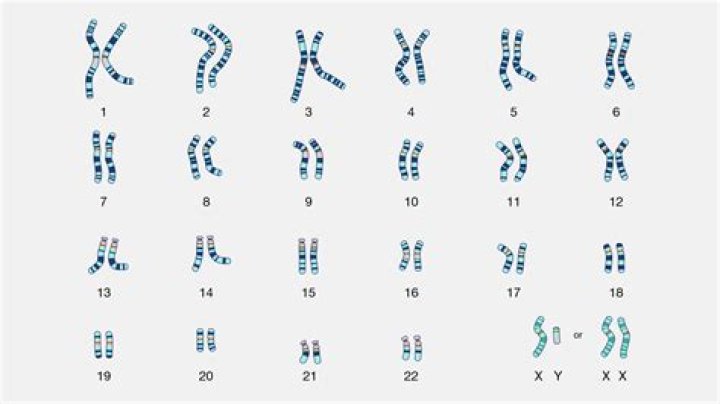
How are karyotypes named?
In a human karyotype, autosomes or “body chromosomes” (all of the non–sex chromosomes) are generally organized in approximate order of size from largest (chromosome 1) to smallest (chromosome 22). … Using this naming system, locations on chromosomes can be described consistently in the scientific literature.
How can the two chromosomes?
How can the two chromosomes that make up a homologous pair differ? They can contain different alleles for the same trait.
How are chromosomes prepared for a karyotype?
Karyotypes are prepared from mitotic cells that have been arrested in the metaphase or prometaphase portion of the cell cycle, when chromosomes assume their most condensed conformations. A variety of tissue types can be used as a source of these cells.
How biologists make a karyotype?
how do biologist make a karyotype? They photograph cells in mitosis, cut out the chromosomes from the photographs, then group the chromosomes together in pairs.
What chromosome is a male?
Each person normally has one pair of sex chromosomes in each cell. The Y chromosome is present in males, who have one X and one Y chromosome, while females have two X chromosomes.
Can you have 43 chromosomes?
A gain or loss in the number of chromosomes from the normal 46 is called aneuploidy. A common form of aneuploidy is trisomy, or the presence of an extra chromosome in cells. “Tri-” is Greek for “three”; people with trisomy have three copies of a particular chromosome in cells instead of the normal two copies.
Is fish a karyotype?
Multifluor FISH generates a karyotype in which each chromosome appears to be painted with a different color. Each “paint” is actually a collection of hybridization probes for sequences that span the length of a particular chromosome.
What can G-banding detect?
G-banding allows each chromosome to be identified by its characteristic banding pattern. The banding pattern can distinguish chromosomal abnormalities or structural rearrangements, such as translocations, deletions, insertions, and inversions.
Is bar a body?
The Barr, or sex chromatin, body is an inactive X chromosome. It appears as a dense, dark-staining spot at the periphery of the nucleus of each somatic cell in the human female.
